Ureters, Urinary Bradder, Urethra Urethra, Urinary Bradder, Urethra Urethra 乌里特拉
章节大纲
-
Communicating with Urine
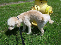
::与尿交流Why do dogs urinate on fire hydrants? Besides “having to go,” they are marking their territory with chemicals in their urine called pheromones . It’s a form of , in which they are “saying” with odors that the yard is theirs and other dogs should stay away. In addition to fire hydrants, dogs may urinate on fence posts, trees, car tires, and many other objects. Urination in dogs, as in people, is usually a voluntary process controlled by the brain . The process of forming urine — which occurs in the — occurs constantly, and is not under voluntary control. What happens to all the urine that forms in the kidneys? It passes from the kidneys through the other organs of the , starting with the ureters .
::狗为什么要在消防水龙头上撒尿? 除了“去去”之外,它们用尿液中的化学物质来标记自己的领土,叫做费洛蒙。 这是一种形式,在这种形式下,它们用气味来“说 ” : 院子是它们的,其他狗应该远离它。 除了消防水龙头,狗也可以在栅栏、树、汽车轮胎和其他许多物体上撒尿。 狗的呼救通常像人一样,是由大脑控制的一种自愿过程。 形成尿的过程 — — 发生在尿液中 — — 经常发生,并且不受自愿控制。 肾脏中的所有尿会怎么样? 它从肾脏通过其他器官,从尿液开始。Ureters
::电流As shown in the figure , ureters are tube-like structures that connect the kidneys with the urinary bladder . They are paired structures, with one ureter for each kidney. In adults, ureters are between 25 and 30 cm (10 to 12 in.) long and about 3 to 4 mm (about 1/8 in.) in diameter.
::如图所示,尿素是连接肾脏和尿囊的类似管状结构,是配对结构,每个肾有一个尿囊,成年人的尿素直径在25至30厘米(10至12英寸)之间,直径约为3至4毫米(约1/8英寸)。Besides the kidneys, the urinary system includes two ureters, the urinary bladder, and the urethra.
::除肾脏外,尿道系统包括两个尿管、尿囊和尿道。Each ureter arises in the pelvis of a kidney (the renal pelvis in the figure ). It then passes down the side of the kidney, and finally enters the back of the bladder . At the entrance to the bladder, the ureters have sphincters that prevent the backflow of urine.
::每个尿素都来自肾脏的骨盆(图中的肾脏骨盆 ) 。 然后,尿素从肾脏的侧面传下去,最后进入膀胱的背部。 在膀胱的入口处,尿素有防止尿液回流的螺栓。Urine collects in the renal pelvis, which is continuous with the ureter. The ureter then carries the urine from the kidney to the urinary bladder.
::尿液在肾脏骨盆中收集,尿素与尿素持续结合,尿素随后将尿液从肾脏运到尿囊。The walls of the ureters are composed of multiple layers of different types of tissues . You can see the layers in the figure . The innermost layer is a special type of epithelium, called transitional epithelium. Unlike the epithelium lining most organs, transitional epithelium is capable of flattening and distending, and does not produce mucus . It lines much of the urinary system, including the renal pelvis, bladder, and much of the urethra , in addition to the ureters. Transitional epithelium allows these organs to stretch and expand as they fill with urine or allow urine to pass through. The next layer of the ureter walls is made up of loose connective tissue containing elastic fibers, nerves , and and lymphatic vessels. After this layer are two layers of smooth muscles , an inner circular layer, and an outer longitudinal layer. The smooth muscle layers can contract in waves of peristalsis to propel urine down the ureters from the kidneys to the urinary bladder. The outermost layer of the ureter walls consists of fibrous tissue.
::尿管的墙壁由不同种类的组织组成的多层组成。 您可以在图中看到这些层。 最深层的一层是特殊类型的上皮类, 称为过渡性上皮。 与大多数器官的上皮类不同, 过渡性上皮类能够拉平和分解, 并且不会产生粘结。 它排出大量尿系统, 包括肾盆、 膀胱和大部分尿道, 除了尿管之外。 过渡性上皮类允许这些器官在填满尿液或允许尿液通过时伸展和膨胀。 下一层尿道壁由松散的连接组织组成, 包含弹性纤维纤维、 神经和淋巴容器。 之后的一层是两层光滑动的肌肉, 内循环层, 外部长度层。 光质的肌肉层可以在透气波中压缩出尿液, 从肾脏到尿囊囊囊。 最外层是由纤维组织组成的。This illustration shows a cross-section of a ureter. The white space inside is the lumen through which urine passes. From the lumen outward, the layers of the ureter wall include transitional epithelium, connective tissue, two layers of muscle fibers, and an outer layer of fibrous tissue.
::这个插图显示了尿素的截面。 里面的白色空间是尿液经过的润滑剂。 从润滑剂向外, 尿素墙的层层包括过渡性肾上腺、 连接组织、 两层肌肉纤维和外层纤维组织。Urinary Bladder
::脉冲推进器The urinary bladder is a hollow, muscular, and stretchy organ that rests on the pelvic floor. It collects and stores urine from the kidneys before the urine is eliminated through urination. As shown in the figure , urine enters the urinary bladder from the ureters through two ureteral openings on either side of the back wall of the bladder. Urine leaves the bladder through a sphincter called the internal urethral sphincter. When the sphincter relaxes and opens, it allows urine to flow out of the bladder and into the urethra.
::尿囊是位于骨盆底部的空洞、肌肉和伸缩器官,在尿液通过尿液消除之前从肾脏中收集和储存尿液,如图所示,尿液从尿囊进入尿囊中,通过膀胱后墙两侧的两个尿道开口进入尿囊中,尿液通过一个称为内尿质螺旋体的螺旋体将膀胱离开。当脊椎放松和打开时,尿液允许尿液从尿囊中流出,进入尿囊中。This diagram of the urinary bladder shows (a) a cross-sectional drawing of the entire bladder and (b) a microscopic cross-section of the tissues in the wall of the bladder.
::本尿囊图显示(a) 整个膀胱的横截面图和(b) 膀胱墙上组织部位的显微剖面图。Like the ureters, the bladder is lined with transitional epithelium, which can flatten out and stretch as needed as the bladder fills with urine. The next layer (lamina propria) is a layer of loose connective tissue, nerves, and blood and lymphatic vessels. This is followed by a submucosa layer, which connects the lining of the bladder with the detrusor muscle in the walls of the bladder. The outer covering of the bladder is peritoneum, which is a smooth layer of epithelial that lines the abdominal cavity and covers most abdominal organs.
::与尿素一样,膀胱与过渡性肾上腺相连接,可以随着膀胱填满尿而粉碎和伸展。下一层(拉米娜阴道)是松散的连接组织、神经、血液和淋巴容器的一层。接着是亚肌肉层,将膀胱的衬里与膀胱壁上的干扰肌肉连接起来。膀胱的外层是腹膜,这是一条光滑的上皮层,连接腹腔腔,覆盖大多数腹部器官。The detrusor muscle in the wall of the bladder is made of smooth muscle fibers controlled by both the autonomic and somatic nervous systems. As the bladder fills, the detrusor muscle automatically relaxes to allow it to hold more urine. When the bladder is about half full, the stretching of the walls triggers the sensation of needing to urinate. When the individual is ready to void, conscious nervous signals cause the detrusor muscle to contract, and the internal urethral sphincter to relax and open. As a result, urine is forcefully expelled out of the bladder and into the urethra.
::膀胱壁上的干扰肌肉是由由自主和体能神经系统控制的光滑肌肉纤维制成的。 膀胱填充时, 干扰肌肉自动放松, 以允许其保持更多的尿液。 当膀胱大约满半时, 墙的伸展触发了需要小便的感觉。 当个人准备消亡时, 有意识的神经信号导致干扰肌肉萎缩, 而内尿素螺栓则放松和开放。 结果, 尿被从膀胱和尿囊中被强行排出, 进入尿囊中 。Urethra
::UrethraThe urethra is a tube that connects the urinary bladder to the external urethral orifice, which is the opening of the urethra on the surface of the body. As shown in the figure , the urethra in males travels through the penis , so it is much longer than the urethra in females. In males, the urethra averages about 20 cm (8 in.) long, whereas in females, it averages only about 4.8 cm (1.9 in.) long. In males, the urethra carries semen (as well as urine), but in females, it carries only urine.
::尿道是一种将尿囊与外部尿囊连接的管子,这是人体表面尿囊的开口,如图所示,雄性尿囊穿透阴茎,比雌性尿囊长得多,雄性尿囊平均约20厘米(8英寸),雌性尿囊平均只有4.8厘米(1.9英寸),雄性尿囊平均只有4.8厘米(1.9英寸)。雄性尿囊携带精液(以及尿液),而雌性尿囊只携带尿液。The urinary bladder and urethra are colored brown to illustrate them in (a) female anatomy and (b) male anatomy. Notice how much longer the male urethra is because it travels through the length of the penis to reach the external urethral orifice.
::尿囊和尿囊是棕色的,用(a) 雌性解剖和(b) 雄性解剖来说明,注意雄性尿囊多长,因为它穿透阴茎进入外尿囊。Like the ureters and bladder, the proximal (closer to the bladder) two-thirds of the urethra are lined with transitional epithelium. The distal (farther from the bladder) third of the urethra is lined with mucus-secreting epithelium. The mucus helps protect the epithelium from urine, which is corrosive. Below the epithelium is loose connective tissue, and below that are layers of smooth muscle that are continuous with the muscle layers of the urinary bladder. When the bladder contracts to forcefully expel urine, the smooth muscle of the urethra relaxes to allow the urine to pass through.
::与尿素和膀胱一样,尿素的三分之二与过渡性肾上腺结合。尿素的三分之一与尿素分泌物(膀胱的距离)相连接。尿素的三分之一与粘结剂分泌物分泌物分泌物分泌物分泌物相连接。粘结剂有助于保护上皮免受腐蚀性尿的侵扰。在上皮下面是松散的连接组织,下面是与尿囊肌肉层相连接的光滑肌肉层。当膀胱与强力排出尿液的合同签订时,尿素的光滑肌肉会放松,让尿液通过。In order for urine to leave the body through the external urethral orifice, the external urethral sphincter (shown in the bladder figure above) must relax and open. This sphincter is a striated muscle that is controlled by the somatic nervous system , so it is under conscious, voluntary control in most people (exceptions are infants , some elderly people, and patients with certain injuries or disorders). The muscle can be held in a contracted state and hold in the urine until the person is ready to urinate. Following urination, the smooth muscle lining the urethra automatically contracts to re-establish muscle tone, and the individual consciously contracts the external urethral sphincter to close the external urethral opening.
::为了让尿液通过外尿道离开身体,外部尿囊(如以上膀胱图所示)必须放松和打开。这个麻痹是受体质神经系统控制的割裂性肌肉,因此在多数人(婴儿、一些老年人和某些受伤或紊乱的病人除外)有意识和自愿控制之下。肌肉可以保持在一个合同状态,并保持尿液,直到人们准备尿便为止。尿尿后,光滑的肌肉将尿囊自动连接起来,以重新建立肌肉音,个人有意识地将外尿质活体连接起来,关闭外尿道开口。Summary
::摘要-
Ureters are tube-like structures that connect the kidneys with the urinary bladder. Each ureter arises at the renal pelvis of a kidney and travels down through the abdomen to the urinary bladder. The walls of the ureter contain smooth muscle that can contract to push urine through the ureter by peristalsis. The walls are lined with transitional epithelium that can expand and stretch.
::尿管是连接肾脏和尿囊的类似管状结构。每根尿囊都出现在肾脏的肾脏骨盆上,从腹部穿到膀胱。尿囊的墙壁有光滑的肌肉,可以通过透透气将尿液排入尿囊。墙壁与可扩展和伸展的过渡性肾上腺相连接。 -
The urinary bladder is a hollow, muscular organ that rests on the pelvic floor. It is also lined with transitional epithelium. The function of the bladder is to collect and store urine from the kidneys before the urine is eliminated through urination. Filling of the bladder triggers the sensation of needing to urinate. When a conscious decision to urinate is made, the detrusor muscle in the bladder wall contracts and forces urine out of the bladder and into the urethra.
::膀胱是位于骨盆地板上的空洞肌肉器官,与过渡性肾上腺相连接;膀胱的作用是在尿液通过尿液消除之前从肾脏中收集和储存尿液;填补膀胱会引发需要尿的感觉;当作出有意识的大小便决定时,膀胱墙上的分泌肌肉会与膀胱和尿尿尿结合,将尿液从膀胱中排出,并排入尿尿囊中。 -
The urethra is a tube that connects the urinary bladder to the external urethral orifice. Somatic nerves control the sphincter at the distal end of the urethra. This allows the opening of the sphincter for urination to be under voluntary control.
::尿道是一种将尿囊与外部尿囊连接的管子。 体能神经控制了尿道阴道端的螺旋体。 这样可以打开用于排尿的螺旋体, 从而在自愿控制下进行排尿。
Review
::回顾1. What are ureters?
::1. 什么是尿素?2. Describe the location of the ureters relative to other urinary tract organs.
::2. 说明尿管相对于其他尿道器官的位置。3. Identify layers in the walls of a ureter. How do they contribute to the ureter’s function?
::3. 辨别尿素墙壁的层层,它们如何促进尿素功能?4. Describe the urinary bladder.
::4. 描述尿囊的情况。5. What is the function of the urinary bladder?
::5. 尿囊的功能是什么?6. How does the nervous system control the urinary bladder?
::6. 神经系统如何控制尿囊?7. What is the urethra?
::7. 什么是尿道?8. How does the nervous system control urination?
::8. 神经系统如何控制小便?9. Identify the sphincters that are located along the pathway from the ureters to the external urethral orifice.
::9. 查明位于从尿素到外部尿道孔道沿线的螺旋体。10. What are two differences between the male and female urethra?
::10. 男女尿道之间有什么区别?11 . True or False: Urine travels through the urinary system due solely to the force of gravity.
::11. 真实或虚假:尿仅仅由于重力而通过尿道系统。12. True or False: Urination refers to the process that occurs starting with the formation of urine in the kidneys, and ending with the elimination of urine from the body.
::12. 真实的或假的:急速是指从肾脏内形成尿液开始,直至从身体中消除尿液的过程。13. When the bladder muscle contracts, the smooth muscle in the walls of the urethra _________ .
::13. 当膀胱肌肉收缩时,尿道墙壁的光滑肌肉。14. Transitional epithelium lines the ______________________.
::14. 过渡性顶部直线a. bladder
::a. 膀胱b. ureters
::b. 尿管c. renal pelvis
::c. 肾骨盆d. all of the above
::d. 以上所有情况Explore More
::探索更多You deposit it in toilets and then you flush and never see it again. Could we be making use of all the pee (and poop) we usually flush away? Watch this TED talk to learn more about the potential use of pee and other human excrement to grow healthier plants and people:
::你把它放在厕所里,然后冲水,再也不会看见它。我们能否利用我们通常冲走的尿尿(和便便)?看TED的演讲,更多地了解尿尿和其他人类粪便的潜在用途,以培养更健康的植物和人:Can holding your pee be bad for you? Learn more here:
::握住你的小便会不会对你不好? -
Ureters are tube-like structures that connect the kidneys with the urinary bladder. Each ureter arises at the renal pelvis of a kidney and travels down through the abdomen to the urinary bladder. The walls of the ureter contain smooth muscle that can contract to push urine through the ureter by peristalsis. The walls are lined with transitional epithelium that can expand and stretch.