显微镜
章节大纲
-
How can we see the details of ?
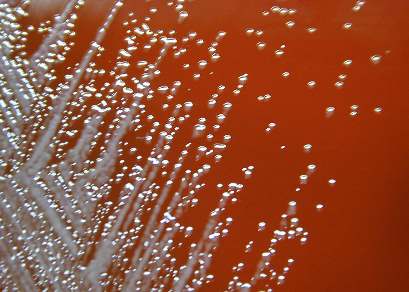
::我们如何看待细节?With the naked eye, bacteria just look like a slimy smear on a petri dish. How can we study them in more detail? The invention of the microscope has allowed us to see bacteria, , and other things too small to be seen with the naked eye.
::以肉眼看,细菌只是看起来像一个粘糊糊糊的涂片 放在花盆上。我们怎样才能更详细地研究它们呢?显微镜的发明让我们可以看到细菌, 和其他太小的东西, 无法用肉眼看出来。The Microscope
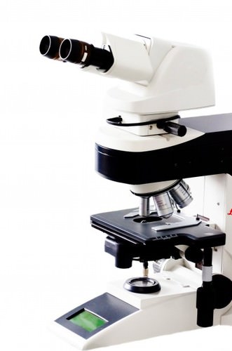
::显微镜Microscopes, tools that you may get to use in your class , are some of the most important tools in biology ( Figure ). A microscope is a tool used to make things that are too small to be seen by the human eye look bigger. Microscopy is the study of small objects using microscopes. Look at your fingertips. Before microscopes were invented in 1595, the smallest things you could see on yourself were the tiny lines in your skin. But what else is hidden in your skin?
::显微镜,你可以在班级中使用的显微镜,是生物学中最重要的工具(图 ) 。显微镜是一种工具,用来制造太小的东西,而人类的眼睛看不到这些小的东西。显微镜是使用显微镜研究小物体。看看你的指尖。在显微镜于1595年发明之前,你能看见的最小的东西是皮肤中的小线。但是,在你的皮肤中还隐藏着什么呢?Basic light microscopes opened up a new world to curious people.
::基本光显微镜为好奇的人开辟了新的世界。Invention of the Microscope
::显微镜的发明Over four hundred years ago, two Dutch spectacle makers, Zacharias Janssen and his son Hans, were experimenting with several lenses in a tube. They discovered that nearby objects appeared greatly enlarged, or magnified . This was the forerunner of the compound microscope and of the telescope.
::四百多年前,两位荷兰美景制作人Zacharias Janssen和他的儿子汉斯正在用一个管子中的几个镜头进行实验,他们发现附近物体看起来大得多或放大。这是复合显微镜和望远镜的前身。In 1665, Robert Hooke, an English natural scientist, used a microscope to zoom in on a piece of cork - the stuff that makes up the stoppers in wine bottles, which is made from tree bark. Inside of cork, he discovered tiny structures, which he called cells. It turns out that cells are the smallest structural unit of living organisms . This finding eventually led to the of the theory that all living things are made of cells. Without microscopes, this discovery would not have been possible, and the would not have been developed.
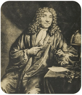
::1665年,英国自然科学家罗伯特·胡克(Robert Hooke)用显微镜放大了一块软木—— 由树皮制成的葡萄酒瓶中的阻塞器。 在软木里,他发现了很小的结构,他称之为细胞。结果发现细胞是活生物体中最小的结构单位。这个发现最终导致一种理论,即所有生物都是由细胞组成的。没有显微镜,这个发现不可能实现,也不会开发出来。Hooke's discovery of the cell set the stage for other scientists to discover other types of organisms. After Hooke, the "father of microscopy," Dutch scientist Antonie van Leeuwenhoek ( Figure ) taught himself to make one of the first microscopes. In one of his early , van Leeuwenhoek took a sample of scum from his own teeth and used his microscope to discover bacteria , the smallest living organism on the planet. Using microscopes, van Leeuwenhoek also discovered one-celled and cells.
::胡克对细胞的发现为其他科学家发现其他类型的生物创造了条件。 胡克(Hooke,“显微镜之父 ” ) 之后,荷兰科学家安东妮·范利乌文豪克(Figure ) 教自己制作第一个显微镜之一。 范利乌文豪克(Van Leeuwenhoek)在早期之一,从自己的牙齿中采集了一批人渣样本,并利用显微镜发现细菌,这是地球上最小的活生物体。凡利乌文豪克(Van Leeuwenhoek)也用显微镜发现了一个细胞和细胞。Today, microscopes are used by all types of scientists, including cell biologists, microbiologists, virologists, forensic scientists, entomologists, taxonomists, and many other types.
::今天,所有类型的科学家都使用显微镜,包括细胞生物学家、微生物学家、病毒学家、法医科学家、昆虫学家、分类学家和许多其他类型的科学家。Antonie van Leeuwenhoek, a Dutch cloth merchant with a passion for microscopy.
::Antonie van Leeuwenhoek, 荷兰服装商, 热衷于显微镜。Types of Microscopes
::显微镜类型Some modern microscopes use light, as Hooke's and van Leeuwenhoek's did. Others may use electron beams or sound waves. Researchers now use these four types of microscopes:
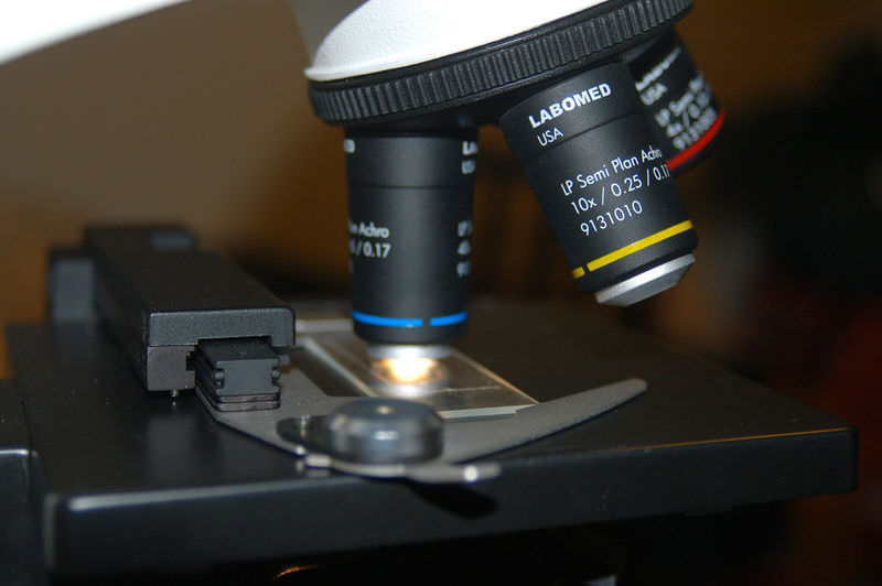
::有些现代显微镜使用光,如胡克和范利乌文豪克所用的光。另一些则使用电子束或声波。研究人员现在使用这四种显微镜:- Light microscopes allow biologists to see small details of a specimen. Most of the microscopes used in schools and laboratories are light microscopes. Light microscopes use lenses, typically made of glass or plastic, to focus light either into the eye, a camera, or some other light detector. The most powerful light microscopes can make images up to 2,000 times larger.
::光显微镜可以让生物学家看到一个标本的小细节。 学校和实验室使用的显微镜大多是光显微镜。 光显微镜使用一般由玻璃或塑料制成的镜片,将光聚焦在眼睛、照相机或其他一些光探测器中。 最强大的光显微镜可以使图像大到2000倍。
- Transmission electron microscopes (TEM) focus a beam of electrons through an object and can make an image up to two million times bigger, with a very clear image.
::传输电子显微镜(TEM)通过一个物体聚焦一束电子,使图像大达200万倍,图像清晰。
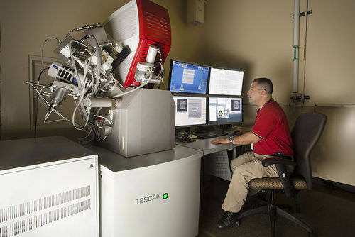
- Scanning electron microscopes (SEM) allow scientists to find the shape and surface texture of extremely small objects, including a paperclip, a bedbug, or even an atom . These microscopes slide a beam of electrons across the surface of a specimen, producing detailed maps of the surface of objects. Magnification in a SEM can be controlled over a range from about 10 to 500,000 times.
::扫描电子显微镜(SEM)使科学家能够发现极小物体的形状和表面纹理,包括纸剪、臭虫甚至原子。这些显微镜将电子束滑过标本表面,绘制物体表面的详细地图。SEM的放大作用可以控制在10至500 000倍之间。
- Scanning acoustic microscopes use sound waves to scan a specimen. These microscopes are useful in biology and medical research.
::扫描声波显微镜使用声波扫描标本,这些显微镜对生物学和医学研究有用。
A scanning electron microscope.
::扫描电子显微镜Science Friday: Stained Glass Conservation
::星期五:染色玻璃保护Stained glass from the Middle Ages is often hundreds of years old. Unfortunately, many of these relics are in need of cleaning and maintenance. In this video by Science Friday, conservator Mary Higgins discusses the methods used to protect the stained glass.
::中世纪的染色玻璃往往有数百年的历史。 不幸的是,许多这些遗迹需要清理和维护。 在科学星期五的录像中,玛丽·希金斯(Mary Higgins)讨论了保护染色玻璃的方法。Summary
::摘要- A microscope is a tool used to make things that are too small to be seen by the naked eye look bigger.
::显微镜是一种工具,用来使肉眼看不到的太小的东西看起来更大。
- Types of microscopes include light microscopes, transmission electron microscopes (TEM), scanning electron microscopes (SEM), and scanning acoustic microscopes.
::显微镜的类型包括光显微镜、传输电子显微镜、扫描电子显微镜和扫描声显微镜。
Explore More
::探索更多Use the resources below to answer the questions that follow.
::利用以下资源回答以下问题。- Using a microscope at (4:01)
::使用显微镜(4:01)
- How should you carry a compound optical microscope?
::你该如何携带复合光学显微镜?
- What procedure should you use when seeking to use the most powerful optical lenses?
::在试图使用最强大的光眼镜时,你应当采用什么程序?
Review
::回顾- What is the purpose of a microscope?
::显微镜的目的是什么?
- What were the findings of Hooke and van Leeuwenhoek?
::Hooke和van Leeuwenhoek有什么发现?
- What are the differences between a light microscope and a scanning electron microscope?
::光显微镜和扫描电子显微镜有什么区别?
- Light microscopes allow biologists to see small details of a specimen. Most of the microscopes used in schools and laboratories are light microscopes. Light microscopes use lenses, typically made of glass or plastic, to focus light either into the eye, a camera, or some other light detector. The most powerful light microscopes can make images up to 2,000 times larger.