16.3 心脏心脏
章节大纲
-
Lub, Dub
::卢布、杜布Lub dub, lub dub, lub dub... That’s how the sound of a beating heart is typically described. Those are also the only two sounds that should be audible when listening to a normal, healthy heart through a stethoscope, as in the photo above. If a doctor hears something different from the normal lub dub sounds, it’s a sign of a possible heart abnormality. What causes the heart to produce the characteristic lub dub sounds? Read on to find out.
::Lub dub, lub dub, lub dub, lub dub bub...这就是跳动心脏的声音通常被描述的方式。这些也是通过听诊器听到正常健康心脏时唯一能听到的两种声音,如上面的照片。如果医生听到不同于正常润滑剂的声音,那就是心脏可能不正常的迹象。是什么原因导致心脏产生典型的润滑剂声音?请继续了解。Introduction to the Heart
::心心入门介绍The heart is a muscular organ behind the sternum (breastbone), slightly to the left of the center of the chest. A normal adult heart is about the size of a fist. The function of the heart is to pump through of the . The continuous flow of blood through the system is necessary to provide all the of the body with oxygen and nutrients , and to remove their metabolic wastes.
::心脏是胸腔(胸骨)后面的肌肉器官,略微靠近胸部中间的左侧。正常的成人心脏与拳头的大小差不多。心脏的作用是抽动。通过这个系统不断流出血液是向所有身体提供氧气和营养,并清除其代谢废料所必须的。Structure of the Heart
::心脏结构The heart has a thick muscular wall that consists of several layers of tissue . Internally, the heart is divided into four chambers through which blood flows. Because of heart valves, blood flows in just one direction through the chambers.
::心脏有一块厚厚的肌肉壁,由几层组织组成。在内部,心脏被分成四间血液流经的室室。由于心脏阀门,血液流向一个方向,通过室室。Heart Wall
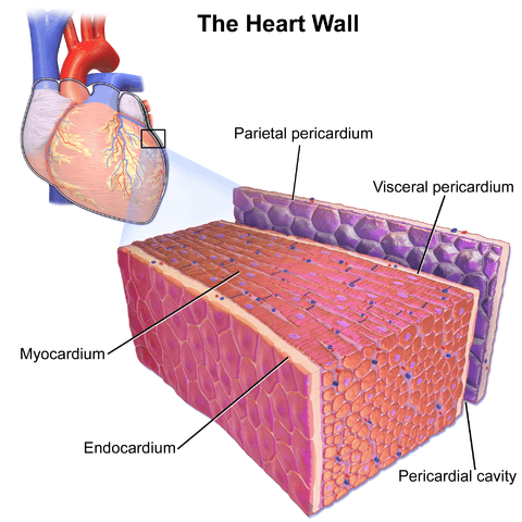
::心脏墙壁As shown in the figure , the wall of the heart is made up of three layers, called the endocardium, myocardium, and pericardium.
::如图所示,心脏的墙由三层组成,称为心内核、心内核和心外核。-
The
endocardium
is the innermost layer of the heart wall. It is made up primarily of simple epithelial cells. It covers the heart chambers and valves. A thin layer of
connective tissue
joins the endocardium to the myocardium.
::心内核是心壁最深层的一层,主要由简单的上皮细胞组成,覆盖心脏室和阀门。一小层连接组织将心内核与心内核连接在一起。 -
The
myocardium
is the middle and thickest layer of the heart wall. It consists of
cardiac muscle
surrounded by a framework of
collagen
. There are two types of cardiac muscle cells in the myocardium: cardiomyocytes — which have the ability to contract easily — and pacemaker cells, which conduct electrical impulses that cause the cardiomyocytes to contract. About 99 percent of cardiac muscle cells are cardiomyocytes, and the remaining one percent is pacemaker cells. The myocardium is supplied with blood vessels and
nerve
fibers via the pericardium.
::心心血管是心脏壁中最厚的一层,由心肌组成,由科伦框架环绕,心肌细胞有两种类型:心心血管细胞——它们能够很容易地结合——和心脏起搏细胞,它们产生电脉冲,导致心血管细胞萎缩,大约99%的心脏肌肉细胞是心血管细胞,其余的1%是心脏起搏细胞。心血管细胞通过腹膜提供血管和神经纤维。 -
The
pericardium
is a protective sac that encloses and protects the heart. The pericardium consists of two membranes (visceral pericardium and parietal pericardium), between which there is a fluid-filled cavity. The fluid helps to cushion the heart, and also lubricates its outer surface.
::是一种保护性囊囊囊,可以包扎并保护心脏。心肌外皮由两个膜组成(心肌外皮和皮肤外皮外皮),介于两个膜之间,中间有一个液体填充的洞穴。液体有助于缓冲心脏,并润滑其外表。
The wall of the heart is made up mainly of myocardium, which consists largely of cardiac muscle.
::心脏的墙壁主要由心心肌组成,心肌主要由心肌组成。Heart Chambers
::心心室As shown in the diagram , the four chambers of the heart include two upper chambers called atria (singular, atrium) , and two lower chambers called ventricles . The atria are also referred to as receiving chambers, because blood coming into the heart first enters these two chambers. The right atrium receives blood from the upper and lower body through the superior vena cava and inferior vena cava , respectively. The left atrium receives blood from the lungs through the pulmonary veins . The ventricles are also referred to as discharging chambers, because blood leaving the heart passes out through these two chambers. The right ventricle discharges blood to the lungs through the pulmonary artery , and the left ventricle discharges blood to the rest of the body through the aorta . The four chambers are separated from each other by dense connective tissue consisting mainly of collagen.
::如图所示,心脏的四个部室包括两个上层室,称为阿提亚( singria, atrium),两个下层室,称为通风室。阿提亚也被称为接收室,因为血液先进入心脏,进入这两个室。右中层通过上部和下部分别通过上部和下部的Vena cava和低等的Vena cava获得血液。左部中层通过肺部通过肺血管获得血液。通风室也被称为释放室,因为留下心脏的血液通过这两个室流出。右心室通过肺动脉向肺中排放血液,左心室通过血管向身体其余部分排放血液。四个部通过主要由钴构成的密集连接组织相互分离。This cross-sectional diagram of the heart shows its four chambers and four valves. The white arrows indicate the direction of blood flow through the heart chambers.
::心脏的横截面图显示了心脏的4个空格和4个阀门。白箭指血流穿过心脏空格的方向。Heart Valves
::心谷The diagram above also shows the location of the heart's four valves. The heart valves allow blood to flow from the atria to the ventricles, and from the ventricles to the pulmonary artery and aorta. The valves are constructed in such a way that blood can flow through them in only one direction, thus preventing the backflow of blood. The four valves are the:
::以上图还显示了心脏四个阀门的位置。心脏阀门允许血液从阿提里亚流到通风室,从通风室流到肺动脉和动脉。阀门的构造方式是,血液只能流到一个方向,从而防止血液回流。四个阀门是:-
tricuspid valve
, which allows blood to flow from the right atrium to the right ventricle
::允许血液从右端端端流到右心室的三端阀门 -
mitral valve
, which allows blood to flow from the left atrium to the left ventricle
::线性阀门,允许血液从左中子流到左心室 -
pulmonary valve
, which allows blood to flow from the right ventricle to the pulmonary artery
::肺阀门,允许血液从右心室流到肺动脉 -
aortic valve
, which allows blood to flow from the left ventricle to the aorta
::允许血液从左心室流到动脉的动脉阀门
Coronary Circulation
::冠状环The cardiomyocytes of the muscular walls of the heart are very active cells, because they are responsible for the constant beating of the heart. These cells need a continuous supply of oxygen and nutrients. The carbon dioxide and waste products they produce also must be continuously removed. The blood vessels that carry blood to and from the heart muscle cells make up the coronary circulation. Note that the blood vessels of the coronary circulation supply heart tissues with blood, and are different from the blood vessels that carry blood to and from the chambers of the heart as part of the general circulation. Coronary arteries supply oxygen-rich blood to the heart muscle cells. Coronary veins remove deoxygenated blood from the heart cells.
::心脏肌肉壁的心血管细胞是非常活跃的细胞,因为它们对心脏不断跳动负有责任,这些细胞需要持续供应氧气和营养素。它们生产的二氧化碳和废物产品也必须不断清除。它们产生的二氧化碳和废物产品也必须不断清除。携带血液和心脏肌肉细胞的血管组成了冠心循环。注意冠状循环的血管用血液供应心脏组织,与作为一般循环的一部分而将血液输送到心脏的血管不同。冠状动脉向心脏肌肉细胞提供含氧血液。冠状血管从心脏细胞中除去脱氧血液。-
There are two coronary arteries — a right coronary artery that supplies the right side of the heart, and a left coronary artery that supplies the left side of the heart. These arteries branch repeatedly into smaller and smaller arteries and finally into capillaries, which exchange gases, nutrients, and waste products with cardiomyocytes.
::有两种冠状动脉,一种是右冠状动脉,提供心脏的右侧,另一种是左冠状动脉,提供心脏的左侧。 这些动脉分支反复进入较小和较小的动脉,最后进入毛动脉,这种动脉交换气体、营养素和含有心血管细胞的废品。 -
At the back of the heart, small cardiac veins drain into larger veins, and finally into the great cardiac vein, which empties into the right atrium. At the front of the heart, small cardiac veins drain directly into the right atrium.
::在心脏的后部,小心脏血管排入大血管,最后排入大心脏血管,再排入右心室。在心脏前部,小心脏血管直接排入右心室。 在心脏前部,小心脏血管排入右心室。
Blood Circulation Through the Heart
::血液通过心脏循环The diagram shows how blood circulates through the chambers of the heart. The right atrium collects blood from two large veins, the superior vena cava (from the upper body) and the inferior vena cava (from the lower body). The blood that collects in the right atrium is pumped through the tricuspid valve into the right ventricle. From the right ventricle, the blood is pumped through the pulmonary valve into the pulmonary artery. The pulmonary artery carries the blood to the lungs, where it enters the pulmonary circulation , gives up carbon dioxide, and picks up oxygen. The oxygenated blood travels back from the lungs through the pulmonary veins (of which there are four), and enters the left atrium of the heart. From the left atrium, the blood is pumped through the mitral valve into the left ventricle. From the left ventricle, the blood is pumped through the aortic valve into the aorta, which subsequently branches into smaller arteries that carry the blood throughout the rest of the body. After passing through capillaries and exchanging substances with cells, the blood returns to the right atrium via the superior vena cava and inferior vena cava, and the process begins anew.
::图表显示血液如何通过心室循环。 右心室从两个大血管中收集血液, 上方的上方的上方的颈部, 和下方的颈部的下方的颈部。 右心室中收集的血液通过三片阀门抽进右心室。 从右心室, 血液通过肺阀抽出进入肺动脉。 肺动脉将血液输送到肺部, 进入肺部循环, 提供二氧化碳, 并收集氧气。 氧化血液从肺部返回肺部, 穿过肺部( 四处), 进入心脏左心室。 从左心室, 血液通过肺阀抽出进入左心室。 从左心室, 血液通过呼吸阀抽进肺部的肺部, 进入肺部, 进入肺循环, 注入二氧化碳, 吸收氧化血液, 从肺部回回回流到血液, 后部, 向下方的脊部, 和下方的血液返回。The flow chart in this diagram summarizes the pathway blood takes as it flows into, through, and out of the heart. Trace the path of blood flow in the diagram of the heart as you follow it through the flow chart.
::此图表中的流程图概述了血液流进、 流出和流出心脏的路径。 在您通过流程图时, 跟踪心脏图中的血液流路径 。Cardiac Cycle
::心脏病周期The cardiac cycle refers to a single complete heartbeat, which includes one iteration of the lub and dub sounds heard through a stethoscope. During the cardiac cycle, the atria and ventricles work in a coordinated fashion so that blood is pumped efficiently through and out of the heart. The cardiac cycle includes two parts, called diastole and systole, which are illustrated in the diagrams .
::心脏循环是指一个完全的心跳,包括一个通过听诊器听到的润滑剂和哑巴音的迭代。在心脏循环期间,腹部和心室以协调的方式工作,以便有效地抽出和抽出血液。心脏循环包括两个部分,即直肠和立体,在图表中说明了这两个部分。-
During
diastole,
the atria contract and pump blood into the ventricles, while the ventricles relax and fill with blood from the atria.
::在氨酸盐期间,阿提里亚合同和将血液抽进腹腔,而腹腔放松,充斥着阿提雅的血液。 -
During
systole,
the atria relax and collect blood from the lungs and body, while the ventricles contract and pump blood out of the heart.
::在疗养院期间,阿提雅放松,从肺部和身体中收集血液,而心血管收缩,抽出心脏的血液。
Diastole is referred to the filling stage, because this is when the ventricles fill with blood. Systole is referred to the pumping stage because this is when the ventricles pump blood out of the heart.
::将丁托尔指向填充阶段, 因为这是脑室充满血液的时候。 质波尔则指向抽泵阶段, 因为这是脑室抽出心脏血的时候 。Electrical Stimulation of the Heart
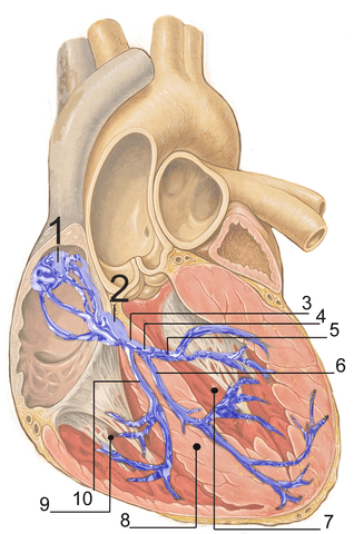
::心脏的电动The normal, rhythmical beating of the heart is called sinus rhythm. It is established by the heart’s pacemaker cells, which are located in an area of the heart called the sinoatrial node (shown in the figure ). The pacemaker cells create electrical signals with the movement of electrolytes (sodium, potassium, and calcium ions) into and out of the cells. For each cardiac cycle, an electrical signal rapidly travels first from the sinoatrial node, to the right and left atria so they contract together. Then, the signal travels to another node, called the atrioventricular node (also shown ), and from there to the right and left ventricles (which also contract together), just a split second after the atria contract.
::心脏正常的、有节奏的跳动被称作关节。它由心脏的心脏起搏器细胞建立,这些细胞位于心脏的一个叫做神经节点的地区(如图所示 ) 。 心脏起搏器细胞随着电解液(钠、钾和钙离子)进出细胞而产生电信号。 对于每个心脏周期,电信号从神经节点到右和左之间快速移动,因此它们相互连接。 然后,信号会飞到另一个节点,称为脑电动节点(也显示在图中 ) , 从那里到左和右心室(也同时连接在一起 ) , 就在亚斯特里亚合同签订后一秒钟。In this drawing of the heart, the numbers refer to (1) the sinoatrial node and (2) the atrioventricular node. The blue lines in the drawing indicate the path of transmission of electrical signals through the heart.
::在绘制心脏时,数字指的是(1) 神经结点和(2) 感应节点。绘图中的蓝线表示通过心脏传输电信号的途径。The normal sinus rhythm of the heart is influenced by the autonomic nervous system through sympathetic and parasympathetic nerves. These nerves arise from two paired cardiovascular centers in the medulla of the brainstem. The parasympathetic nerves act to decrease the heart rate, and the sympathetic nerves act to increase the heart rate. Parasympathetic input normally predominates. Without it, the pacemaker cells of the heart would generate a resting heart rate of about 100 beats per minute, instead of a normal resting heart rate of about 72 beats per minute. The cardiovascular centers receive input from receptors throughout the body, and act through the sympathetic nerves to increase the heart rate, as needed. I ncreased physical activity, for example, is detected by receptors in muscles, , and tendons . These receptors send to the cardiovascular centers, causing sympathetic nerves to increase the heart rate, and allowing more blood to flow to the muscles.
::心脏正常的鼻音节奏受自主神经系统通过同情和同情性寄生神经的影响。 这些神经来自脑电图中两对相配的心血管中心。 寄生虫病神经会降低心率, 同情性神经会增加心率。 偏执性输入通常占主导地位。 没有它, 心脏的心脏起搏器细胞将产生大约每分钟100节的休眠心率, 而不是每分钟大约72节的正常休眠心率。 心血管中心会收到全身体受体的感应器的输入, 并按需要通过同情性神经来增加心跳率。 例如, 增加的体能活动是通过肌肉、 和 管的感应器被检测到的。 这些感应器会传到心血管中心, 导致心动神经增加心跳, 并允许更多的血液流向肌肉。Besides the autonomic nervous system, other factors can also affect the heart rate. For example, thyroid and adrenal hormones (such as epinephrine) can stimulate the heart to beat faster. The heart rate also increases when drops or the body is dehydrated or overheated. On the other hand, cooling of the body and relaxation — among other factors — can contribute to a decrease in the heart rate.
::除了自主神经系统之外,其他因素也可能影响心率,例如甲状腺和肾上腺激素(如肾上腺素)可以刺激心脏跳得更快,当坠落或身体脱水或过热时,心率也会上升,另一方面,身体冷却和放松——除其他因素外——会促使心率下降。Feature: Human Biology in the News
::特著:《新闻》中的人类生物学When a patient’s heart is too diseased or damaged to sustain life, a heart transplant is likely to be the only long-term solution. The first successful heart transplant was undertaken in South Africa in 1967. Over the past two decades in the United States, about 2,400 hearts were transplanted annually . The problem is that far too few hearts are available for transplant, and many patients die each year waiting for a life-saving heart to become available.
::当病人的心脏受到太多疾病或损伤而无法维持生命时,心脏移植可能是唯一的长期解决方案。 第一次成功的心脏移植是1967年在南非进行的。 在过去20年中,美国每年约有2,400颗心脏移植。 问题在于用于移植的心脏太少,每年许多病人死亡,等待救命心脏的出现。Hearts for transplant have to be used within four hours of the death of the donor. In addition, the hearts can only come from brain-dead individuals whose hearts are removed while they are still healthy. Then, the hearts are placed on ice inside picnic coolers to be transported to a waiting recipient. The four-hour window means that traffic jams, bad weather , or other unforeseen delays often result in a heart being in less than optimal condition by the time it arrives at its destination. Unfortunately, there is no way to know if the heart will start up again after it is transplanted until it is actually placed in the recipient’s body. In up to seven percent of cases, a transplanted heart does not work, and has to be removed.
::用于移植的心脏必须在捐赠者死亡后四小时内使用。 此外,心脏只能来自那些在身体健康时心脏被摘除的脑死的人。 之后,心脏被放在野餐冷却器中的冰上,以便运送给等待接收者。 4小时窗口意味着交通堵塞、恶劣天气或其他意外的延误往往导致心脏在到达目的地时处于不理想状态。 不幸的是,在心脏被移植之后,直到移植到接收者的身体时,还无法知道心脏是否会再次发作。 最多7%的病例中,移植心脏不会起作用,必须移除。A medical device company in Massachusetts called TransMedic was featured in many news stories when it developed the Organ Care System, commonly referred to as “heart-in-a-box.” The system takes a new approach to maintaining donated hearts until they are transplanted. The box is heated and contains a device that pumps oxygenated blood through the heart while it is being transported to the recipient. This extends the time the heart can remain healthy and usable by up to 12 hours. It also allows the heart to be monitored, so it is kept in optimal condition while en route . The end result, ideally, is that the recipient gets a healthier heart with less chance of failure for the new organ, and a lower risk of death.
::在马萨诸塞州,一家名为TransMedic的医疗设备公司在很多新闻报道中被报道了,当时它开发了有机器官护理系统,通常被称为“心箱中”系统。 该系统采取了一种新的方法来维持捐赠的心脏直到移植为止。 盒子是加热的,里面装着一个在将心脏运到接收者时通过心脏抽吸氧血的装置。这延长了心脏保持健康和可用的时间,最长可达12小时。它也允许对心脏进行监测,因此在行进途中将心脏保持最佳状态。 最终的结果最好是接受者得到更健康的心脏,而新器官的失灵机率更低,死亡风险也更低。As of mid-2016, the heart-in-a-box system had already been used for several successful heart transplants in other countries. At that time, the system was also undergoing clinical trials in the United States to assess its effectiveness in promoting positive recipient outcomes. Developers of the heart-in-a-box predict that the system could increase the number of usable donor hearts by as much as 30 percent, thus greatly increasing the number of patients who are saved from death by heart failure.
::截至2016年年中,在其他国家,心脏在箱中系统已经用于几次成功的心脏移植。 当时,该系统还在美国进行临床试验,以评估其在促进积极接收效果方面的成效。 箱中心脏开发者预测,该系统可以将可用捐赠者心脏数量增加30%,从而大大增加了因心脏衰竭而免于死亡的患者人数。Summary
::摘要-
The heart is a muscular organ behind the sternum and slightly to the left of the center of the chest. Its function is to pump blood through the blood vessels of the cardiovascular system.
::心脏是胸骨后面的肌肉器官,在胸口中间的左侧略微靠近,其功能是通过心血管系统的血管抽血。 -
The wall of the heart consists of three layers. The middle layer, the myocardium, is the thickest layer and consists mainly of cardiac muscle. The interior of the heart consists of four chambers, with an upper atrium and lower ventricle on each side of the heart. Blood enters the heart through the atria, which pump it to the ventricles. Then the ventricles pump blood out of the heart. Four valves in the heart keep blood flowing in the correct direction and prevent backflow.
::心脏的壁由三层组成。 中间层, 心肌, 是最厚的层, 主要由心脏肌肉组成。 心脏的内部由四间室组成, 心脏的两侧都有上端的中心和下层的心室。 血从心脏进入心脏, 将心脏泵到心室。 然后, 心室抽出血液。 心脏的四扇阀门保持血液向正确的方向流动, 防止回流 。 -
The coronary circulation consists of blood vessels that carry blood to and from the heart muscle cells, and is different from the general circulation of blood through the heart chambers. There are two coronary arteries that supply the two sides of the heart with oxygenated blood. Cardiac veins drain deoxygenated blood back into the heart.
::冠状循环由血管组成,它们携带血液进入和流出心脏肌肉细胞,与通过心脏室的血液一般循环不同,有两种冠状动脉为心脏两侧提供氧化血液,心血管将脱氧血液排入心脏。 -
Deoxygenated blood flows into the right atrium through veins from the upper and lower body (superior and inferior vena cava, respectively), and oxygenated blood flows into the left atrium through four pulmonary veins from the lungs. Each atrium pumps the blood to the ventricle below it. From the right ventricle, deoxygenated blood is pumped to the lungs through the two pulmonary arteries. From the left ventricle, oxygenated blood is pumped to the rest of the body through the aorta.
::脱氧血液通过上部和下部的血管(上部和下部的雄性大肠杆菌)流进右中子中子,氧血液通过肺部的4个肺血管流进左中子中子,每个将血液泵到下部的通风室。从右心室,脱氧血液通过两道肺动脉抽入肺部。从左心室,氧血液通过动脉抽到身体其余部分。 -
The cardiac cycle refers to a single complete heartbeat. It includes diastole — when the atria contract — and systole, when the ventricles contract.
::心脏循环是指一个完全的心跳,包括直肠-当阿提里亚合同时的直肠-和直肠-直肠-直肠-直肠-直肠-直肠-直肠。 -
The normal, rhythmic beating of the heart is called sinus rhythm. It is established by the heart’s pacemaker cells in the sinoatrial node. Electrical signals from the pacemaker cells travel to the atria, and cause them to contract. Then, the signals travel to the atrioventricular node and from there to the ventricles, causing them to contract. Electrical stimulation from the autonomic nervous system and hormones from the endocrine system can also influence heartbeat.
::心脏的正常、有节奏跳动被称为鼻弦节奏。它由心脏心脏起搏细胞在神经结节中建立。起搏细胞的电讯从心脏起搏细胞传到阿提雅,并导致它们收缩。然后,信号传到动脉结点,从那里传到心室,导致它们收缩。来自自动神经神经系统以及内分泌系统的荷尔蒙的电动刺激也可以影响心跳。
Review
::回顾1. What is the heart, where is located, and what is its function?
::1. 什么是心脏,在哪里,其作用是什么?2. Outline the structure of the heart.
::2. 概述心脏结构。3. Describe the coronary circulation.
::3. 描述冠状动脉循环。4. Summarize how blood flows into, through, and out of the heart.
::4. 概述血液如何流进、流进和流出心脏。5. Define the cardiac cycle. Identify its two parts.
::5. 界定心脏循环,标明其两部分。6. Explain what controls the beating of the heart.
::6. 解释控制心脏跳动的是什么。7. What are the two types of cardiac muscle cells in the myocardium? What are the differences between these two types of cells?
::7. 心肌内两种心肌细胞是什么类型?这两种细胞之间有什么区别?8. Match each of the three layers of the walls of the heart (endocardium, myocardium, and pericardium) with the description that best matches it below.
::8. 将心脏的三层壁(心电图、心电图、心电图和心电图)各与以下最符合的描述相匹配。a. protects the heart
::a. 保护心脏b. covers the heart valves
::b. 覆盖心脏阀门c. responsible for the beating of the heart
::c. 对心脏跳动负责9. Is the blood that flows through the mitral valve oxygenated or deoxygenated? Explain your reasoning.
::9. 流经线性阀门的血液是否氧化或脱氧?解释一下你的推理。10. True or False: The coronary arteries carry blood to the heart.
::10. 真实或假:冠状动脉将血液带入心脏。11. True or False: Systole is when the heart is contracting, and diastole is when the heart is fully relaxed.
::11. 真实或假:症候是心脏萎缩时的症状,不眠是心脏完全放松时的症状。12. Explain why the blood from the cardiac veins empties into the right atrium of the heart. Focus on function (rather than anatomy) in your answer.
::12. 解释为何心脏血管的血液会排入心脏的右心部,在回答时注重功能(而不是解剖学)。Explore More
::探索更多More women than men die of heart disease, but heart research has long focused on men. In the following TED talk, pioneering doctor C. Noel Bairey Merz shares what we know and don't know about women's heart health, including the very different heart attack symptoms women experience, and why doctors often miss them.
::女性死于心脏病的人数多于男性,但心脏研究长期以来一直以男性为主。 在接下来的TED演讲中,开创性博士C. Noel Bairey Merz分享了我们对女性心脏健康的了解和不了解,包括女性心脏病发作症状的不同,以及为什么医生常常想念她们的原因。Check out this video to see how heart valve replacement surgery is carried out:
::看看如何进行心脏阀门更换手术: -
The
endocardium
is the innermost layer of the heart wall. It is made up primarily of simple epithelial cells. It covers the heart chambers and valves. A thin layer of
connective tissue
joins the endocardium to the myocardium.